


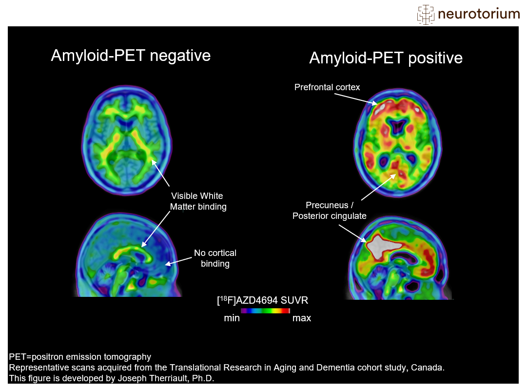
Examples of amyloid-negative (left) and amyloid-positive (right) positron emission tomography (PET) scans. In this figure, the amyloid-PET ligand [18F]AZD4694 is shown. The hotter colours indicate more amyloid-PET tracer binding, and colder colours indicate low amyloid-PET tracer binding. The prototypical amyloid-positive PET pattern is of elevated amyloid in the brain’s posterior cingulate cortex, prefrontal cortex, as well as lateral parietal and temporal cortices. These regions display minimal tracer uptake in the amyloid-negative case (left), who has low binding across the whole cerebral cortex. Finally, it is important to note that amyloid-PET tracers have some binding to white matter, which explains the small amounts of hotter colours in the amyloid-negative case.
More information on amyloid-PET visual reads can be found in Johnson KA, Minoshima S, Bohnen NI, et al. “Appropriate use criteria for amyloid PET: a report of the Amyloid Imaging Task Force, the Society of Nuclear Medicine and Molecular Imaging, and the Alzheimer’s Association”. Alzheimers Dement. 2013;54(3):476-490
Related content


Brain drug delivery systems, allied with their characteristics for overcoming the blood-brain barrier (BBB).

The use of a single treatment in heterogeneous populations has proved problematic in clinical trials undertaken in patients with neurodegenerative diseases.